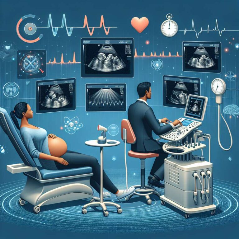
Day and colleagues ran a randomized controlled trial to test whether Artificial Intelligence assistance could improve the efficiency of fetal ultrasound screening for congenital heart disease without compromising diagnostic performance. Sonographers were assigned to perform scans with Artificial Intelligence support or the standard method, and outcomes included detection accuracy, scan and reporting time, and cognitive load. The trial concluded that Artificial Intelligence-assisted scans achieved similar sensitivity and specificity to standard scans, while examinations were completed more quickly and with lower reported workload.
The study enrolled 59 sonographers randomized 1:1 to intervention and control groups. All sonographers received standardized training, followed a study-specific protocol, and were blinded to the clinical status of pregnant participants. On each trial day, one sonographer per group scanned three participants, comprising two with healthy fetuses and one with a fetus with congenital heart disease. Primary outcomes were sensitivity and specificity for congenital heart disease detection, and secondary outcomes included the total time to complete the scan and report as well as the sonographers’ cognitive load measured by the NASA-TLX survey. The evidence rating for the study was 1, indicating excellent quality.
Artificial Intelligence assistance did not produce statistically significant differences in sensitivity or specificity for detecting congenital heart disease or all fetal structural malformations compared with the standard approach. The between-group differences for congenital heart disease detection were 3.8 percent for sensitivity and 7.7 percent for specificity, with 97.5 percent confidence intervals crossing zero. Efficiency improved with Artificial Intelligence support, as the median scan duration was shorter by 9.3 minutes with a 95 percent confidence interval of 7.4 to 11.1. Cognitive load was also reduced in the intervention group, with a mean difference in NASA-TLX score of 10.0 and a 95 percent confidence interval of 4.6 to 15.4.
Authors noted limitations, including self-selection of sonographers that may limit generalizability, potential awareness that a malformation was present despite blinding to specific diagnoses, and the possibility of heightened caution in a small research setting. Even with these constraints, the findings provide encouraging evidence that Artificial Intelligence can streamline fetal anomaly ultrasound workflows by reducing scan duration and cognitive burden, without degrading diagnostic performance.

